(To see other currencies, click on price)
MORE ABOUT THIS BOOK
Main description:
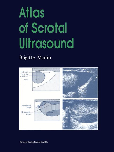
This unique ultrasound atlas is dedicated entirely to pathology of the scrotal contents. The 357 illustrations constitute the major proportion of the book, each of them is accompanied by a labelled diagram and detailed description. The atlas covers the essential embryology and ultrasound anatomy, the principal clinical presentations encountered in practice by the clinician and radiologists, and the differential diagnosis of intrascrotal cysts and calcifications. For each chapter there is a short but precise text, supplemented by tables or decision-making algorithms followed by the relevant images.
Contents:
Embryology; technical notes and ultrasound anatomy.- The acute scrotum.- The chronically enlarged scrotum.- Impalpable testicular tumours within a clinically normal scrotal sac.- Male infertility.- Post-surgery scrotal sac.- Calcifications of structures within the scrotum and the periscrotal region.- Cystic structures of the scrotal contents.- Alphabetical index.
PRODUCT DETAILS
Publisher: Springer (Springer-Verlag Berlin and Heidelberg GmbH & Co. K)
Publication date: August, 2014
Pages: 200
Weight: 543g
Availability: Available
Subcategories: Radiology, Urology