(To see other currencies, click on price)
MORE ABOUT THIS BOOK
Contents:
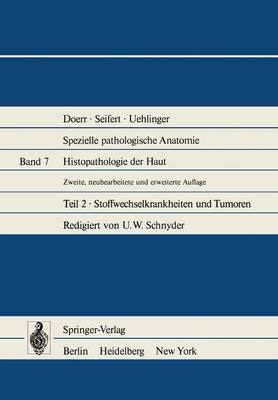
Stoffwechselkrankheiten der Haut.- A. Kohlenhydratstoffwechsel.- I. Diabetes mellitus 1..- 1. Allgemeines.- 2. Gefassveranderungen beim Diabetes mellitus.- II. Glykogenspeicherkrankheiten.- B. Fettstoffwechsel.- Morphologische Befunde bei den Xanthomen.- I. Familiare Lipoproteinmangelzustande.- II. Primare Hyperlipoproteinamien.- 1. Hyperlipoproteinamie Typ I.- 2. Hyperlipoproteinamie Typ II.- 3. Hyperlipoproteinamie Typ III.- 4. Hyperlipoproteinamie Typ IV.- 5. Hyperlipoproteinamie Typ V.- III. Sekundare Hyperlipoproteinamien.- IV. Lipidosen.- 1. Sphingomyelinose (= Morbus Niemann-Pick).- 2. Glucocerebrosidose (= Morbus Gaucher).- 3. Sulfatidlipidosen (= metachromatische Lipodystrophie).- 4. Ceramidtrihexosidose (= Angiokeratoma corporis diffusum universale Fabry).- Anhang: Hyalinosis cutis et mucosae Urbach-Wiethe.- C. Eiweissstoffwechsel.- I. Antikoerpermangelsyndrom.- II. Paraproteinamien.- 1. Plasmocytom.- 2. Makroglobulinamie Waldenstroem.- 3. "Heavy Chain Disease".- 4. Kryoglobulinamien.- III. Amyloidosen.- 1. Primare cutane Amyloidosen.- 2. Sekundare cutane Amyloidosen.- Anhang: Ochronose (Alkaptonurie).- D. Purin- und Pyrimidinstoffwechsel.- Die Gicht.- E. Porphyrinstoffwechsel.- I. Symptomatische cutane hepatische Porphyrie.- II. Porphyria variegata.- III. Porphyria congenita Gunther.- IV. Erythrohepatische Protoporphyrie.- F. Calciumstoffwechsel.- I. Dystrophische Verkalkungen der Haut.- II. Metastatische Verkalkungen der Haut.- G. Pigmentstoffwechsel (exkl. Melanin).- I. Argyrose der Haut.- II. Hamosiderinablagerungen in der Haut.- H. Cutane Mucinosen.- I. Hautveranderungen bei Funktionsstoerungen der Schilddruse.- 1. Hypothyreose.- 2. Hyperthyreose.- II. Cutane Mucinosen bei normaler Schilddrusenfunktion.- 1. Skleromyxoedem Arndt-Gottron und Lichen myxoedematosus.- 2. Scleroedema (adultorum) Buschke.- 3. Mucinosis follicularis (= Alopecia mucinosa).- J. Mucopolysaccharidosen.- Literatur.- Tumoren der Haut.- A. Epidermoide Tumoren.- I. Epidermale Naevi.- 1. Epithelialer Schleimhautnaevus.- 2. Epidermoidcysten.- 3. Seborrhoische Keratose.- 4. Akrotrichom (Inverted Follicular Keratosis).- 5. Formenkreis des sog. intraepidermalen Epithelioms.- 6. Klarzellenacanthom.- 7. Keratoacanthom.- 8. Warziges Dyskeratom.- 9. Benigne Stachelzellkeratose.- II. Pramaligne epidermoide Lasionen.- 1. Actinische Keratosen.- 2. Sonstige epidermale Pracancerosen.- a) Keratosen durch ionisierende Strahlen.- b) Sog. Teerkeratosen.- c) Arsenkeratosen.- 3. Morbus Bowen.- 4. Erythroplasie Queyrat.- 5. Pracanceroese orale Leukoplakien.- 6. Pracanceroese Cheilopathie.- III. Maligne epidermoide Tumoren.- 1. Plattenepithelcarcinom.- a) Acantholytisches Plattenepithelcarcinom.- b) Bowen-Carcinom.- 2. Low-grade Malignancy Carcinoma.- a) Verrucous Carcinoma.- b) Genitales Riesencondylom (Buschke-Loewenstein-Tumoren).- B. Adnexoide Hauttumoren.- I. Naevi und Tumoren des Haarfollikels.- 1. Naevus pilo-follicularis (N.p.-f.).- 2. Trichofolliculom.- 3. Tricholemmom.- 4. Tricho-Adenom.- 5. Pilomatrixom.- 6. Tricho-Epitheliom.- 7. Maligne Tumoren des Haarfollikels.- Pilar Tumor of the Scalp.- II. Naevi und Tumoren der Talgdrusen.- 1. Naevus sebaceus (N.s.).- 2. Talgdrusen-Hyperplasie.- 3. Talgdrusen-Adenom.- 4. Talgdrusen-Epitheliom.- 5. Talgdrusen-Carcinom.- III. Cysten und cystische Tumoren des Haar-Talgdrusenkomplexes.- 1. Naevus comedonicus.- 2. Retentionscyste.- 3. Steatocystoma multiplex (S.m.).- 4. Dermoidcyste..- 5. Tricholemmcyste.- IV. Naevi und Tumoren der Schweissdrusen (Formenkreis der Hidradenome).- 1. Naevus sudoriparus.- 2. Sog. dermales Cylindrom.- 3. Hidradenome mit vorwiegend ekkrinen Merkmalen.- a) Ekkrines Porom.- b) Ekkrines Spiradenom.- c) Klarzellen-Hidradenom.- d) Chondroides Hidradenom.- e) Syringom.- f) Maligne ekkrine Hidradenome.- ?) Schweissdrusencarcinom (im engeren Sinn).- ss) Malignes ekkrines Porom.- y) Malignes Klarzellen-Hidradenom.- 4. Hidrocystome.- 5. Hidradenome mit vorwiegend apokrinen Merkmalen.- a) Hidradenoma papilliferum.- b) Syringocystadenoma papilliferum.- c) Maligne apokrine Hidradenome.- V. Tumoren der Ceruminaldrusen.- VI. Morbus Paget.- 1. Mammarer Morbus Paget.- 2. Extramammarer Morbus Paget.- C. Basaliome.- 1. Nodulares bzw. nodulo-ulceroeses Basaliom.- 2. Pigmentiertes Basaliom.- 3. Oberflachliches Basaliom.- 4. Destruierendes Basaliom.- 5. Sklerodermiformes Basaliom.- 6. sog. pramalignes Fibroepitheliom.- 7. Naevoide Basaliome (bzw. Basaliomatosen).- 8. Basalzellcarcinom und sog. metatypisches Epitheliom.- D. Fehl- und Neubildungen des melanocytaren Systems.- I. Formenkreis des Naevuszell-Naevus (NZN).- 1. Junktionaler Naevuszell-Naevus.- 2. Epidermo-dermaler Naevuszell-Naevus ("Compound-Naevus").- 3. Dermaler Naevuszell-Naevus.- 4. Blasenzell-Naevus.- 5. Halo-Naevus.- 6. Congenitaler Tierfellnaevus.- 7. Benignes juveniles Melanom.- 8. Blauer Naevus.- II. Melanoma in situ.- 1. Lentigo maligna (Hutchinson-Dubreuihl).- 2. Pagetoides Melanoma in situ.- III. Melanoma malignum.- 1. Lentigo Maligna Melanoma (LMM).- 2. Superficial Spreading Melanoma (SSM).- 3. Primary Nodular Melanoma (NM).- 4. Maligner Blauer Naevus (MBN).- E. Neurogene Hauttumoren.- I. Nasales "Gliom".- II. Ganglioneurom.- 1. Ganglioneuroblastom.- 2. Neuroblastoma sympathicum.- III. "Echtes Neurom".- 1. Traumatisches,,Neurom".- IV. Tumoren der Nervenhullzellen.- 1. Neurilemmom.- 2. Neurofibrom (Neurofibromatosis v. Recklinghausen).- 3. Neurofibrosarkom.- 4. Meningeom.- V. Granulares Neurom.- VI. Malignant Granular Cell Tumor.- F. Mesenchymale Hauttumoren.- I. Tumoren des Bindegewebes.- 1. Bindegewebsnaevus.- Fibrous Hamartoma of Infancy.- 2. Dermale Fibrome.- a) Fibrome der papillaren Dermis.- b) Fibrome der reticularen Dermis.- c) Perifolliculare Fibrome (p.F.).- 3. Keloid.- 4. Histiocytom (Dermatofibrom).- Juveniles Xanthogranulom (j.X.).- 5. Cutanes Myxofibrom.- Cutane Schleimcyste.- 6. Pseudo-Sarkome.- a) Infantile digitale Fibromatose.- b) Fasciitis nodularis pseudosarcomatosa (F.ps.).- c) Desmoidtumor.- d) Atypisches Fibroxanthom.- 7. Sarkome der Haut.- a) Dermatofibrosarcoma protuberans.- b) (Subcutane) Fibrosarkome.- II. Tumoren des Fettgewebes.- 1. Naevus lipomatodes superficialis.- 2. Embryonales Lipoblastom.- 3. Lipom.- 4. Hibernom.- 5. Liposarkome.- III. Tumoren der Muskulatur.- 1. Cutane Leiomyome.- 2. Cutanes Leiomyosarkom.- 3. Cutanes Rhabdomyosarkom.- IV. Tumoren mit Knorpel- oder Knochendifferenzierung.- 1. Cutanes Chondrom.- 2. Cutanes Chondrosarkom.- 3. Cutane Osteosis ("Osteom").- V. Synoviale Tumoren.- 1. Benignes (riesenzelliges) Synovi(al)om.- 2. Malignes Synovi(al)om.- Literatur.- Fehl- und Neubildungen der Blut- und Lymphgefasse.- A. Angiektatische Naevi.- I. Congenitale Naevi teleangiectatici (Naevi flammei).- 1. Naevi teleangiectatici mediales et symmetrici.- 2. Naevi teleangiectatici laterales (Naevi flammei).- 3. Naevi flammei laterales mit assoziierten Fehlbildungen.- II. Tardive angiektatische Naevi und Fehlbildungen.- 1. Diffuse genuine Phlebangiektasie (Phlebarteriektasie).- 2. Angiektasien (,"senile Angiome") des freien Lippenrandes (Pasini).- 3. Cirsoides Aneurysma.- 4. Venous Lake.- 5. Naevi aranei (Gefassspinnen, Spidernaevi).- 6. Angiectasia racemosa ("Angioma" racemosum).- 7. Teleangiectasia haemorrhagica hereditaria (Rendu-Osler).- 8. Angiectasia serpiginosa ("Angioma serpiginosum Hutchinson").- B. Angiokeratotische Naevi.- I. Angiokeratome.- 1. Angiokeratoma acroashycticum digitorum (Mibelli).- 2. Angiokeratoma punctiforme scroti s. vulvae (Fordyce-Sutton).- 3. Angiokeratoma corporis diffusum (Fabry).- 4. Angiokeratoma circumscriptum corporis naeviforme.- 5. Angiectasia eruptiva thrombotica, solitares, papuloeses Angiokeratom.- II. Angioma verrucosum.- III. Lymphangiokeratome.- 1. Lymphangioma circumscriptum naeviforme.- 2. Lymphangioma circumscriptum localisatum, Lymphangiokeratom, Lymphangiectasia eruptiva, capillares Aneurysma der Lymphgefasse.- C. Angiomatoese Naevi und Neubildungen.- I. Capillare Angiome.- 1. Planotuberoese und tuberonodoese Hamangiome.- 2. Kasabach-Merritt-Syndrom.- 3. Multiloculare Hamangiomatose des Sauglingsalters.- 4. Progressive multiple Angiome (Darier).- 5. Tardive ("senile") Angiome, "Teleangiectasia papulosa disseminata".- 6. Eruptives Angiom, "Granuloma" teleangiectaticum, "Granuloma" pediculatum s. "pyogenicum".- 7. Angiolymphoide Hyperplasie mit Eosinophilie (Summerly-Wells), papulare Angioplasie, "atypisches Granuloma pediculatum".- 8. Gemmangiom (Orsos).- 9. Juveniles Hamangioendotheliom.- II. Cavernoese Angiome und benigne Neubildungen der Gefasswande..- 1. Arterielle Cavernome (cavernoese Hamangiome mit arterieller Differenzierung.- 2. Venoese Cavernome (cavernoese Hamangiome mit venoeser Differenzierung).- 3. Blue Rubber Bleb Nevus Syndrom.- 4. Mafucci-Syndrom (Cavernome mit Dyschondroplasie).- 5. Angioleiomyom.- 6. Hamangiopericytom (Stout-Murray).- 7. Lymphangioma cavernosum.- D. Glomustumoren (Angiomyoneurome, Glomangiome).- I. Isolierte Glomustumoren.- II. Multiple systematisierte Glomustumoren (systematisierte Glomangiomatose).- III. Multiple disseminierte und familiare Glomustumoren (hereditare Glomangiomatose).- IV. Acraler arteriovenoeser Tumor.- E. Maligne Tumoren der Blut- und Lymphgefasse.- I. Sarcoma idiopathicum haemorrhagicum multiplex (Kaposi).- II. Stewart-Treves-Syndrom (Lymphangiosarkom bei Lymphoedem).- III. Hamangioendothelioma malignum, angioplastisches Sarkom.- IV. Angioplastisches Reticulosarkom.- V. Hamangiosarkome.- VI. Lymphangiosarcoma (Lymphangioendothelioma).- VII. Angio-Endotheliomatosis proliferans systematisata.- Literatur.- Lymphoreticulare Hyperplasien und Neoplasien der Haut.- A. Allgemeiner Teil.- I. Einleitung.- Begriffsbestimmungen.- II. Cellulare Bestandteile der Cutis.- 1. Lymphatische Zellen.- a) Lymphocyten.- b) Immunoblasten.- c) Plasmazellen.- d) Weitere lymphatische Zellen.- 2. Monocyten - Histiocyten - Makrophagen.- 3. Reticulumzellen.- a) Die histiocytische oder phagocytische Reticulumzelle.- b) Die fibroblastische Reticulumzelle oder faserassoziierte Reticulumzelle.- c) Die dendritische Reticulumzelle.- d) Die interdigitierende Reticulumzelle.- 4. Mastzellen.- III. Pathogenese cutaner Lymphome.- IV. Diagnose cutaner Lymphome.- V. Diagnostische Methoden.- B. Spezieller Teil.- I. Lymphoreticulare Hyperplasien der Haut (Cutane Pseudolymphome).- 1. Benigne cutane Lymphoplasien.- a) Lymphadenosis benigna cutis.- b) Lymphocytare Infiltrationen bestimmter Art.- ?) Lymphocytic Infiltration of the Skin.- ss) "Palpable Migratory Arciform Erythema".- y) Eruptive disseminierte lymphocytare Infiltrationen.- c) Besondere Arzneireaktionen.- d) Persistierende Arthropoden-Reaktionen.- 2. Lymphomatoide Papulose.- 3. Actinisches Reticuloid.- 4. Rundzellerythematose; reticulare erythematoese Mucinose; REM-Syndrom.- 5. Angiolymphoide Hyperplasie mit Eosinophilie.- 6. Angio-immunoblastische Lymphadenopathie (Lymphogranulomatosis X).- II. Lymphoreticulare Neoplasien der Haut (Cutane Lymphome).- 1. Non-Hodgkin-Lymphome.- Klinisch-pathologische Korrelationen cutaner Non-Hodgkin-Lymphome.- Klinisches Bild der Hautveranderungen bei Non-Hodgkin-Lymphomen.- Klassifikation cutaner Non-Hodgkin-Lymphome.- Cutane Non-Hodgkin-Lymphome mit niedrigem Malignitatsgrad.- a) Lymphocytische Lymphome.- ?) Chronische lymphatische Leukamie (CLL).- ss) Mycosis fungoides.- y) Sezary-Syndrom.- Anhang: Sezary-Syndrom und Lymphknotenbefall.- ?) Pagetoide Reticulose - Woringer-Koloppsche Krankheit.- ?) Haarzeil-Leukamie (,"hairy cell leukemia").- b) Immunocytome.- Anhang: Plasmocytom (multiples Myelom; plasmocytisches Lymphom).- c) Centrocytisches Lymphom.- d) Centroblastisch-centrocytisches Lymphom.- Cutane Non-Hodgkin-Lymphome mit hohem Malignitatsgrad.- a) Centroblastisches Lymphom.- b) Immunoblastisches Lymphom.- c) Lymphoblastische Lymphome.- d) Burkitt-Lymphom.- 2. Morbus Hodgkin (Lymphogranulomatose Paltauf-Sternberg.- III. Maligne (reticulo-) histiocytare Erkrankungen der Haut (Erkrankungen des Monocyten-Makrophagen-Histiocyten-Systems).- 1. Maligne Histiocytose.- 2. Reticulosarkom.- 3. Reticulosen.- 4. Histiocytosis X.- a) Letterer-Siwesche Krankheit.- b) Hand-Schuller-Christiansche Krankheit.- c) Eosinophiles Granulom.- Anhang: Lymphomatoide Granulomatose.- Literatur.- Myeloproliferative Erkrankungen der Haut.- A. Allgemeiner Teil.- B. Spezieller Teil.- I. Varianten der myeloischen Leukamie.- 1. Eoesinophilenleukamie.- 2. Basophilenleukamie.- II. Monocytenleukamien und myelo-monocytare Leukamien.- Literatur.- Mastzellenkrankheiten.- Mastocytose und maligne Mastzellenreticulose.- I. Allgemeines.- II. Klassifikation.- III. Cutane Mastocytosen.- 1. Isoliertes Mastocytom.- 2. Urticaria pigmentosa (U.p.).- 3. Teleangiectasia macularis eruptiva perstans.- 4. Diffuse (erythrodermatische) cutane Mastocytose.- Symptome der Histamin- und Heparinwirkung.- IV. Mastocytose als Systemkrankheit.- a) Hautveranderungen.- b) Lymphknoten und Skeletveranderungen.- V. Maligne Mastzellenreticulose.- Literatur.
PRODUCT DETAILS
Publisher: Springer (Springer-Verlag Berlin and Heidelberg GmbH & Co. K)
Publication date: January, 2012
Pages: None
Weight: 914g
Availability: Available
Subcategories: General Practice
From the same series